Most patients will not need a dental MRI
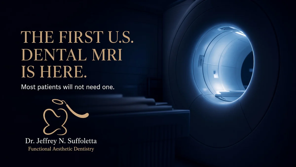
On March 9, 2026, Dentsply Sirona and Siemens Healthineers said the U.S. Food and Drug Administration had cleared the first dental-dedicated MRI system in the United States: the MAGNETOM Free.Max Dental Edition.
That sentence will travel farther than the machine will. Magnetic resonance imaging is not a new idea in medicine. What is new is a scanner built around jaws, teeth, nerves, and joints instead of a borrowed hospital head coil and a 45-minute slot in radiology. The companies say a supporting clinical trial found uses in endodontics, periodontics, TMJ evaluation, tooth extraction, and orthodontics. They also say the average workflow can stay under 20 minutes, and that the field of view is limited to dental structures so brain tissue is not in the picture.
They are equally clear about where the box is meant to live, hospitals, large clinics, and universities, not every general practice with a panoramic unit in a closet.
What X-rays see, and what they miss
Everyday dentistry runs on ionizing radiation. Bitewings find cavities between back teeth. A panoramic film maps wisdom teeth and jaw joints in two dimensions. Cone-beam CT (CBCT) adds a 3-D bone map before implants or impacted-tooth surgery. Those tools are fast, familiar, and good at hard tissue like enamel, dentin, cortical bone.
They are weaker at soft tissue. A disc in the temporomandibular joint, a swollen ligament, the difference between active inflammation and old scar, the vitality of a pulp, or the exact course of a nerve next to a buried third molar can be guesswork on a radiograph. Dentists infer those things from pain tests, probing, and experience. Sometimes they guess wrong, or they order another CBCT and add dose.
MRI does not use X-rays. It aligns hydrogen in water and fat with a magnetic field and reads the signal as those nuclei relax. Fluid-rich tissue lights up. That is why medical MRI already owns much of joint, disc, and nerve imaging. Dentistry stayed away for practical reasons: scanners were expensive, rooms were shielded vaults, exams were long, metal restorations distorted the picture, and few radiologists spoke “tooth.”
What “dental-dedicated” is supposed to fix
The Free.Max Dental Edition is a variant of Siemens’ compact 0.55-tesla Free.Max platform—far below the 1.5T and 3T magnets common in hospitals. Lower field strength usually means a weaker signal. The trade the companies are selling is a wider, more installable magnet, fewer siting headaches, better behavior around some metal, and software that reconstructs a usable dental image anyway. A dedicated dental coil sits close to the face. Specialized software keeps the field of view on teeth and jaws.
Dentsply Sirona’s imaging line already includes systems such as Axeos, Orthophos SL, and Heliodent Plus. The MRI is meant to sit beside those machines, not replace them. Andreas Schneck, head of magnetic resonance at Siemens Healthineers, has framed the project as opening a new clinical area rather than stealing work from existing dental X-ray.
Development involved scientists in Europe and the United States. Aarhus University’s Prof. Rubens Spin-Neto led clinical-trial work used for European CE validation, reported in 2025. Klinikum Dortmund in Germany was described as the first dental clinic to install the system. In the United States, the University of Minnesota School of Dentistry had already partnered on an early research scanner and later hosted hands-on training. A ClinicalTrials.gov listing (NCT06948162) describes an observational study there comparing ddMRI with routine dental images and medical TMJ MRIs.
What the companies say the trial showed
Company materials list three capabilities again and again.
- First, the scanner can separate active inflammation from healthy or scar tissue. That matters in periodontics and around dying roots, where a dark spot on a radiograph can be old healing or a live problem.
- Second, it can offer a noninvasive read on pulp vitality, whether the tissue inside the tooth still has a fluid signal—alongside the cold tests and electric pulp tests already used in the chair.
- Third, it can show teeth relative to neighbors and nerves, including structures such as the lingual nerve that matter before a difficult extraction.
Those are manufacturer claims tied to a validation trial, not a decade of community-practice outcomes. A separate 2026 feasibility paper in the International Endodontic Journal used the same 0.55T Dental Edition with a seven-channel coil on a small series: 18 teeth in nine patients who already had recent CBCT. Observers could see root tips, periapical bone, and lamina dura in every case. Agreement with clinical vitality testing was substantial (kappa 0.77, accuracy 0.88). Agreement on apical periodontitis against the reference workup was higher (kappa 0.87, accuracy 0.94). Scan time in that protocol was about 18 minutes per tooth. That is encouraging. It is not a substitute for a large, independent diagnostic-accuracy trial in ordinary offices.
Who might be sent for one
A referral is easiest to justify when soft tissue is the question and extra radiation is a real cost.
TMJ patients with clicking, locking, or pain that does not match a panoramic film are the obvious group. Medical TMJ MRI already exists; a dental-dedicated unit is an attempt to make that exam shorter and more tooth-aware.
Endodontic cases in which vitality tests conflict, or a CBCT lucency will not declare itself as active disease versus scar, are another. Periodontal and peri-implant inflammation that looks similar to burned-out infection on X-ray is a third.
Impacted wisdom teeth sitting against the inferior alveolar canal, and some orthodontic or surgical plans that need nerve and disc context without stacking more CBCT dose—especially in children who may be imaged more than once—fit the radiation argument.
A cracked filling, a routine cavity, a simple crown prep, or a hygiene exam does not. Bitewings will remain the workhorse. A dental MRI will not find interproximal enamel decay the way a good bitewing does. Bone detail on CBCT is still the standard map for many implant sites.
Who should not assume they are a candidate
MRI is not a spa treatment. Ferromagnetic implants, certain older aneurysm clips, some cochlear devices, and loose metal around the face can be unsafe or can wreck the image. Many modern dental implants and orthodontic appliances are not automatic disqualifiers, and lower field strength is often kinder to metal than 3T, but screening is mandatory. Claustrophobia is common. Contrast dye, when a medical MRI uses it, is a separate decision; dental protocols described so far emphasize anatomy and fluid, not a trip through an IV.
Cost and geography will sort patients faster than physics. A 0.55T magnet is cheaper than a flagship hospital 3T. It is still a capital project with shielding, safety zones, trained staff, and a radiologist or dentist who can read the study. Until that network exists outside a handful of centers, “FDA-cleared” will mean “available somewhere,” not “available Tuesday at the strip-mall office.”
How to read the clearance
FDA clearance of a magnetic resonance diagnostic device is a 510(k) finding of substantial equivalence for a defined use. It is not a mandate that every toothache get an MRI. It is not proof that ddMRI outperforms CBCT for bone, or that it should be billed as a screening tool.
The honest sentence for patients is narrower. Dentistry now has a radiation-free, soft-tissue scanner designed for the mouth, cleared for U.S. sale, aimed at complex cases in large institutions. If a dentist mentions one, the follow-up questions write themselves: What question will this scan answer that a radiograph or CBCT cannot? Who will read it? What does it cost, and will insurance treat it as medical MRI or as dental imaging? Are there metal or medical reasons to skip it?
Most people in a dental chair this year will still leave with a small rectangular film or a cone-beam volume, not a magnet. The news is that when the problem is a disc, a nerve, or an inflammation X-rays cannot name, there is finally a dental-specific MRI on the American market to ask. That is a real expansion of the toolkit. It is not the new checkup.
Related Article:
- Why the ADA is Calling Out Dental Insurance
- Why We’re Doubling Down on New Oral Cancer Standards
- Do You Really Need a Whole-Body Dentist?
- Sleep-Focused Dentistry
We look forward to seeing you at your next visit! If you need to schedule an appointment or have a question, give us a call today.