The Advantages of High Tech X-rays
In contemporary dental practice, one sure way of making a precise diagnosis is the use of the high tech digital x-rays, also described as digital dental radiographs. These are x-rays that use digital sensors to produce detailed images of your teeth and gum while displaying any medical issues affecting your dental structure. Because digital dental x-rays are extremely detailed, your dental expert can easily assess your oral structures, make an apt diagnosis, and draw a suitable treatment plan and follow-up.
How digital dental radiography works
Traditional film-based x-rays use a photographic film to capture a dental image. Instead, digital x-rays use a special sensor to capture images and display them for analysis on a digital device, such as a computer. The images can be captured with a direct method where the sensor is placed inside the mouth to capture images of a targeted oral structure.
In the indirect method, a scanner converts film-based x-rays to digital images. The semi-indirect method integrates the sensor and scanner to transform x-rays into a digital film.
RELATED ARTICLE: Advantages of Advanced Dental Technology
Types of digital dental x-rays
Digital dental x-rays can be classified as intraoral when the sensor is placed inside the mouth or extraoral when the dental image is generated from outside the mouth.
702-671-0001
Keep Your Teeth Healthy, Ask us About Our In-House Dental Benefits

Intraoral x-rays
Intraoral x-rays are precise and provide precise details of the targeted oral structure. They can define details on cavities, the tooth bone, and any dental conditions undetectable to the naked eye. Intraoral x-rays can be:
- Bitewing X-rays: the patient bites the film to allow imaging of the lower and upper teeth at a specific point of the mouth. The image captures the tooth from the crown to the supporting bone, aiding the diagnosis of dental structure and the effects of gum disease on the tooth’s bone.
- Periapical X-rays: these x-rays focus on the periapical area of the tooth (the area surrounding the roots). They image the entire tooth from the crown to the root by focusing on the upper or the lower jaw at a time. The dentist can easily detect advanced periodontitis and endodontic abscesses from these radiographs.
- Occlusal X-rays: these capture images of the entire arc of the jaw. They show details on the position of teeth and are key in monitoring tooth development especially in children.
RELATED ARTICLE: Waterlase Dentistry Provides Gentle and Fast Service to Patients
Extraoral X-Rays
While they may not have the precision of intraoral x-rays, extraoral digital dental images are opportune for a complete image of your dental structure. They can take one of these forms:
- Panoramic X-rays: the digital x-ray machine rotates around the patient to generate a complete image of the oral structure. Because of their completeness, panoramic x-rays allow a comprehensive treatment plan for all the detected oral conditions.
- Cephalometric projections: these target the patient’s entire head in view of examining the dental and surrounding structures. They are especially common with orthodontist working on teeth alignment.
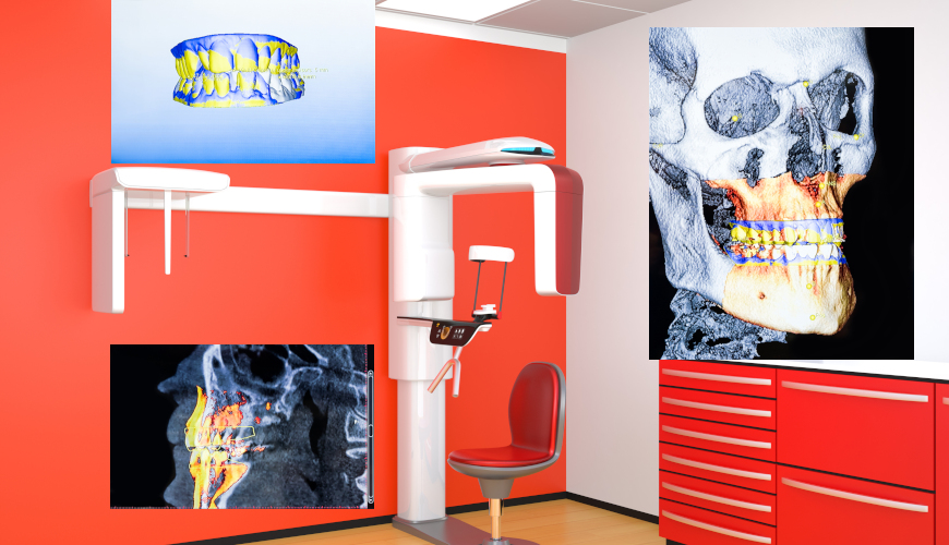
- Multi-slice computed tomography: they focus on a specific part of the mouth while blocking all other parts. The core purpose of using these radiographs is to detect hard-to-see dental structures and focus on details.
- Cone beam computerized tomography: they produce 3D images that focus on teeth and surrounding structures. The dentist can use them as a guide when performing complex dental procedures.
- Sialography: entails the injection of a radiopaque dye into the salivary gland to create a contrast medium allowing the dentist to take an x-ray known as a sialogram. The image is used to detect the functionality of the salivary glands and rule out salivary gland pathologies in manifesting oral or facial inflammations.
RELATED ARTICLE: Las Vegas Dentist is Leading the Way with Advanced Dental Technology
Advantages of High Tech Digital X-Rays
The precision of all these high tech digital x-rays comes with several advantages.
- The x-rays are captured by a sensor in the patient’s mouth and delivered immediately to the digital device. This makes them more efficient than the typical x-rays where time is needed to process the film.
- The digital images offer a more accurate analysis because they can be zoomed to create a contrast.
- The dentist can store digital copies of the radiographs and so keep a record of the patient’s progress.
- Digital x-rays expose the patient to lesser amounts of radiation, hence, adhering to the ‘As Low As Reasonably Achievable’ radiation exposure principle.
- When patients are referred to other specialists such as an oncology doctor in cases of oral cancer, digital radiographs can be easily transferred between practitioners.
Because digital radiography is relatively new in dentistry, dental practitioners are required to observe the dental practice code which commits them to train in new equipment and techniques.
Call 702-671-0001 or visit Functional Aesthetic Dentistry online to learn more.